124 | The complete guide to urinary tract endoscopy for veterinary nurses
We’re finishing our endoscopy series today with a less common, but by no means less important, procedure - lower urinary tract endoscopy, AKA cystoscopy.
This is a relatively uncommon procedure - I’d never seen one outside of referral practice - but just like our other endoscopic procedures, there are tons of ways we can put our nursing skills to good use, and do more to support patients during urinary investigations.
So in this episode that’s exactly what we’ll do. Over the next half an hour or so, we’ll look at what urinary endoscopy actually involves, the common indications for it, how to prepare, handle and maintain the equipment we use, and - most importantly - the nursing considerations when supporting your urinary patients.
So which areas of the urinary tract do we scope, and how do we do it?
If you’ve been here for a while, then you’ll know that I always like to revisit our anatomy and physiology (I promise it’ll be brief!) before we get stuck into the specific disease or procedure.
It gives us a much better understanding of what we’re doing and how we do it - and cystoscopy is no exception to this. You’ll see that the kit we use is very different from patient to patient, and this depends almost entirely on their anatomy - so stick with me for the next few minutes, and everything else will make a lot more sense as we go on.
Cystoscopy, aka urethrocystoscopy or urinary endoscopy, is the endoscopic examination of the lower urinary tract.
In females (who make up the majority of the patients we perform this in), this consists of:
The vaginal vestibule
The vagina
The urethral opening
The urethra
The urinary bladder
The ureteral openings - the points where the two ureters enter the bladder.
The bladder consists of the apex (the ‘top’ or cranial aspect of the bladder), the body (the main portion of the bladder), and the neck (the caudal end of the bladder, which narrows and funnels into the urethra). The ureters should enter the bladder at a region called the trigone, which is a flat, triangular section near the bladder neck.
The anatomy of the rest of the lower urinary tract - the urethra, especially - is entirely dependent on the species and sex you’re scoping.
In a female dog, the urethra is relatively short, wide, and straight, so we can use a shorter, rigid endoscope for the procedure.
However, in males, the anatomy is completely different, which means the kit we use needs to be different, too. Because the urethra is long, curved, and runs through the os penis, you physically can't pass a rigid scope along it, so male dogs need a flexible scope instead. This scope needs to not only be flexible, but also narrow enough to pass through a narrow (typically 3- 4 mm) opening.
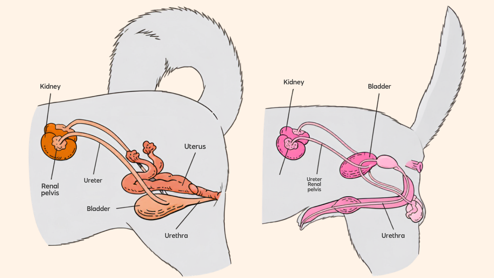
Anatomy of the female (left) and male (right) canine urogenital system.
Ok, so that’s the anatomy covered - how is a cystoscopy performed?
Firstly, the procedure needs to be performed clean-to-aseptically. The urinary tract is a clean environment, and we do not want to introduce contamination and cause a UTI. This means the endoscope needs to be sterile (or cold-soaked if you do not have an autoclavable scope or access to EO), the procedure should be performed in a clean environment, and the endoscopist should be wearing sterile gloves.
After induction, the prepuce or perivulval area is clipped and aseptically prepared as you would do for urinary catheterisation, and the scope is introduced. The urethra (+/- vestibule and vagina, if you’re scoping a female patient) is examined, before the scope is advanced into the urinary bladder.
In the bladder, the urine is drained and replaced with warmed sterile saline. This firstly distends the bladder, allowing thorough assessment of the area, and secondly aids visualisation by replacing urine with clear liquid.
Inside the bladder, the mucosa is examined for evidence of masses, erosions, inflammation or other abnormalities, and the presence of any stones is noted.
It’s also really important that the clinician locates and visualises each ureteral opening - and stays at each one long enough to see a jet of urine exit the ureter and enter the bladder.
After visual assessment of each area, tissue samples are collected as needed - for things like culture as well as histopathology. Depending on the reason for your patient’s cystoscopy (which we’ll come to in a second), we may also need to perform other interventional procedures, such as laser treatment of ectopic ureters or lithotripsy (breaking up) stones.
At the end of the procedure, the bladder is drained, and the patient recovers. They may have some local discomfort (cystitis-type pain) but can typically go home within 24-48 hours, depending on the individual.
So that’s what a cystoscopy is - what are the indications for one in dogs and cats with lower urinary abnormalities?
Like the other scopes we've covered across this series, cystoscopy tends to be performed late in our diagnostic approach to urinary disease. It isn’t a common procedure in general - especially in male dogs (I’ve seen 2 in my entire career!) - and it tends to be reserved for patients where other imaging/diagnostics have not identified the problem.
In most cases, patients have already had routine bloodwork, multiple urinalyses and cultures, and imaging - such as ultrasound and/or contrast radiographs - before a scope is performed. These may have failed to identify the issue, or the patient may not have responded to appropriate treatment as expected.
Patients who undergo urethrocystoscopy usually have:
Chronic or recurrent urinary tract infections that aren't resolving
Persistent or recurrent haematuria
Dysuria or stranguria - difficulty and straining to urinate
Pollakiuria - passing small amounts frequently
Urinary incontinence, particularly in younger female dogs (which is suspicious for ectopic ureters)
Suspected uroliths, or stones spotted incidentally on imaging
Suspected masses in the bladder, urethra or vagina
Trauma to the lower urinary tract
Anatomical abnormalities, like ectopic ureters or vestibulovaginal remnants
Abnormal findings on imaging, urinalysis or urine sediment, such as neoplastic cells
The two most common scenarios are urinary incontinence in a young dog and haematuria or stranguria in an older dog.
In our young female dogs who are leaking urine (usually since a puppy), we're looking for an ectopic ureter - a ureter that enters in an abnormal location, meaning the urine bypasses the normal sphincters and leaks out. Cystoscopy is the ideal method for finding and defining these, because we can directly visualise them. Often, we’ll correct the abnormality under the same procedure with laser ablation - opening up the division between the ureter and bladder, allowing urine to enter the bladder more normally.
In older dogs with persistent haematuria or straining, we're usually worried about a mass. Urothelial carcinoma, aka transitional cell carcinoma, is one we see most commonly, and cystoscopy lets the clinician see how far that mass extends and take a guided biopsy to confirm it.
What about cats?
Cystoscopy isn’t generally performed in cats - mostly due to their size. In most cats, especially male cats, the urethra is just too narrow for a scope to fit.
That being said, most feline lower urinary disorders don’t actually need cystoscopy to diagnose or correct them, so in most cases, it isn’t too much of a problem.
What kit do we need to perform cystoscopy, and how is this kit managed?
We’ve already spoken a little about the different scopes needed for male and female cystoscopy, but let’s look at the kit in more detail - and talk about how each is handled, prepared, and maintained, since that is a big part of our role as nurses and technicians.
Firstly, the type of scope we use depends entirely on the patient we’re scoping.
We scope our female dogs (and some female cats, though rarely) using a rigid telescope. This is a 30-degree rigid telescope with a working sheath, exactly like we use for rhinoscopy. You can actually use your rhinoscope to do this; just make sure you sterilise it before use, and handle it aseptically.
The male dogs need a flexible urethrocystoscope, again sterilised before use (or cold-disinfected if EO is not available). In some cases, a narrow bronchoscope may be small enough to fit into the urethra - but a cystoscope is preferred.
These are usually 2.5- 3 mm in diameter, long enough to travel along the entire urethra and into the bladder, and have an instrument channel for biopsy collection. However, because the scope is narrow, the instrument channel is small, and any biopsies we collect are often < 1 mm in size, which can limit the diagnostic information we get from them.
Our scopes will be connected to the endoscopy tower, camera, and light cable as needed - most flexible scopes will plug directly into the tower without needing an external camera or light source, but rigid telescopes will need to be attached to a camera head and light cable.
Alongside the scope, we need to think about ancillary kit.
This will include our biopsy forceps, grasping forceps or stone retrieval baskets, sterile saline and giving sets, and potentially, a diode laser and laser fibre for interventional procedures.
And of course, as veterinary nurses and technicians, a big part of our role is preparing, handling and maintaining all of this equipment.
This involves visually inspecting everything, leak-testing the cystoscope, checking functions before the patient is anaesthetised, and ensuring the biopsy forceps and other ancillary kit are functional and undamaged.
After the procedure, the items should be cleaned (even just a quick pre-clean) as soon as possible.
As we discussed earlier in the series, dried fluid, blood, and tissue can block the endoscope incredibly quickly, and in a scope this narrow, this is even more important.
After the procedure, do a quick patient-side clean to prevent obstruction by brushing and flushing the channels, and then you can set the kit safely aside, giving you a bit more time before a ‘proper’ clean is performed.
That full clean is the same as wepp discussed for our rhinoscope and bronchoscope: we clean the kit in a neutral enzymatic cleaning solution, brushing and flushing through the channels and gently wiping the scope body itself. After soaking for the required duration, we repeat the process in a high-level instrument disinfectant, then rinse the scopes in distilled water.
Once dried, they are sterilised - ideally using ethylene oxide, since the rapid heating and cooling cycles in most autoclaves can damage even autoclavable rigid scopes.
For storage, the rigid telescopes and their instruments should ideally live in purpose-made boxes or trays that stop them from being squashed or knocked. Flexible scopes should be stored hanging vertically in a closed cupboard, not in their boxes, where the tubing is coiled for long periods.
What about anaesthetic management during cystoscopy? How can we nurse these patients in the periop period?
Cystoscopy is always performed under general anaesthesia, and this means a lot of scope for us to support these patients.
There is no particular anaesthetic protocol these patients need, so our job is to collaborate with our vet team to create a multimodal, tailored anaesthetic plan for each individual patient and monitor them much the same way as we would any other anaesthetised patient.
That being said, there are a few specific considerations:
Antibiotics are usually administered around the time of the procedure, in the same manner as for a surgical procedure, as we’re introducing instruments into the urinary tract.
During the procedure, we need to monitor carefully for bladder over-distension. If the saline continues to run as the bladder fills, there’s a risk of urinary rupture. Monitor bladder size carefully, and alert the vet if you feel it is becoming too full.
And - although this isn’t a true anaesthetic consideration - we need to collect urine samples BEFORE we scope. If the patient hasn’t had a urinalysis recently, one should be collected before we introduce any fluids or collect any samples, as these will change our results.
Aside from anaesthetic management, how else can we support cystoscopy patients as veterinary nurses?
There are a few other things to consider with this procedure, including preparation, fluid management, cleanliness, and sample collection.
First, preparation.
As I mentioned, this is a clean procedure. It should happen early in the procedure list, in a clean environment, with sterile equipment.
Second, we need to look at fluid management.
We know the bladder needs to be clear and full for cystoscopy. Once the clinician has introduced the scope into the bladder, we usually manage that. We’ll use a large syringe and 3-way tap to empty the bladder, then refill it carefully with saline, avoiding overdistension.
Pressure infusers should be avoided, and the fluid flow should be turned down as needed to avoid overfilling the bladder.
When filling the bladder, we want to use warm saline to prevent hypothermia. However, it can be helpful to have a bag of cold saline on hand as well - especially if there’s haemorrhage, because that cold saline can be manually squirted against that mucosa to try and stop the bleeding.
The bladder mucosa is delicate and can be friable, especially in a patient with lower urinary inflammation or neoplasia - so some bleeding from the biopsy sites is to be expected.
We also need to collect that fluid as it drains out. This is a very wet procedure, and patients can easily become soaked if not careful - increasing their risk of hypothermia further. I recommend using something like a tub table (if you have one in a clean area), or failing that, using a fluid collection drape to catch the fluid and keep your patient dry. Wicking bedding should be placed on top of these, to avoid the patient lying against a wet drape.
And then we also need to talk about sampling.
Just like our other endoscopic procedures, sample collection is really important. How we collect, handle, and store these directly affects the pathologist’s interpretation and, therefore, the information we get back from the lab (and the answers we get for our patient).
In cystoscopy, we're taking several samples for histopathology, with at least one biopsy set aside in sterile saline for culture and sensitivity.
As nurses and technicians, we often handle the instruments, collect these samples for the vet, and submit them for analysis.
So here are a few top tips to help with this:
First, advance any forceps gradually, roughly a centimetre at a time, and stop if you feel any resistance. In male dogs, this is especially common because we’re often introducing forceps through a bent scope, which can increase the risk of channel damage.
Second, keep your instruments closed until you can see that the tip has fully exited the channel or sheath, to avoid channel damage from opening them prematurely. Most forceps have a small ‘wing’ or ‘wire’ underneath the cups that also opens when the forceps open, and if this is too close to the channel, it can cause damage - so make sure there’s enough room.
When advised to by the vet, push purposefully but carefully against the area being sampled, close the forceps firmly, and then withdraw them fully in the closed position.
To free the sample from the forceps, shake or rinse it into a sterile pot of saline rather than teasing it off with a needle - because a needle can damage both the tissue and the forceps.
And then after the procedure, place your culture sample (usually the smallest/worst-quality sample) in a small saline pot (labelled ‘culture’) and the rest of the samples in formalin. Then send them, along with a summary of the history, imaging findings (where applicable) and details of any current or recent medications - especially antibiotics. This is particularly important, because many patients will have received multiple courses of different antibiotics before the procedure.
Our nursing care then continues into recovery.
Once the procedure's finished, these patients are monitored closely until they're fully recovered from their anaesthetic, and there are a few specific things we need to look out for in recovery.
The first is urination.
It’s really important that we see these patients passing urine, ideally with a good stream, before they are discharged. There's a risk of urethral swelling, inflammation, and blood clots (which in severe cases could cause obstruction) - so we want to make sure our patient can urinate comfortably before they go home.
Next is pain.
These patients can be uncomfortable around the area after the procedure, so we need to monitor pain scores and administer appropriate analgesia under veterinary direction.
We also need to prevent interference. These patients are often uncomfortable around the area that has been clipped and cleaned, as well as their urethra/bladder, which means that self-trauma is pretty common. This can easily cause further inflammation and infection, so an Elizabethan collar is indicated in recovery.
Aside from this, the rest of our considerations are similar to any other patient.
We also need to talk to our clients about what to monitor for, what to expect in ongoing recovery at home, and when they should return to see us - as well as any other specific considerations based on the individual patient’s needs.
So that’s an overview of urethrocystoscopy in dogs - when and why it is performed, how, and the role we play as veterinary nurses and technicians.
To sum up everything we’ve discussed this episode, cystoscopy is the direct endoscopic examination of the lower urinary tract - the vestibule, vagina, urethra, bladder and the ureteral openings at the trigone.
It’s performed when our workup so far has not revealed a specific cause, or when appropriate medical management hasn’t resolved the patient’s signs.
Usually, it’s performed in patients with recurrent infections, haematuria, straining, incontinence, masses or anatomical abnormalities. And although it’s relatively uncommon overall, it’s almost never performed in cats - so in most cases, it is our dogs we’ll be supporting through this.
Our nursing role is vast and includes selecting, preparing, and managing the appropriate equipment; creating tailored anaesthetic plans under veterinary direction and prescription; and continuing that care throughout and after the procedure.
From aseptic set-up, positioning, site preparation, kit management, preventing bladder overdistension, helping manage bleeding, and monitoring urination in recovery (and more!), there are so many ways we can use our skills to support these patients.
Did you enjoy this episode? If so, I’d love to hear what you think. Take a screenshot and tag me on Instagram (@vetinternalmedicinenursing) so I can give you a shout-out and share it with a colleague who’d find it helpful!
Thanks for learning with me this week, and I’ll see you next time!
References and Further Reading
Cox, S. (2009). Overview of small animal urinary tract endoscopy. DVM360, Available at: https://www.dvm360.com/view/overview-small-animal-urinary-tract-endoscopy-proceedings (Accessed 17th August 2026)
Lhermette, P. (2015). Urethrocystoscopy of the urinary system in dogs and cats. In Practice, 37(9), pp. 445-455. Available at: https://doi.org/10.1136/inp.h4796 (Accessed 17th August 2026)
Lhermette, P., Sobel, D. (2008). Rigid endoscopy: urethrocystoscopy and vaginoscopy. In: BSAVA Manual of Canine and Feline Endoscopy and Endosurgery. BSAVA, Quedgeley, pp. 142-157.
Llido, M., Vachon, C., Dickinson, M., Beauchamp, G., & Dunn, M. (2020). Transurethral cystoscopy in dogs with recurrent urinary tract infections: Retrospective study (2011-2018). Journal of veterinary internal medicine, 34(2), 790–796. Available at: https://doi.org/10.1111/jvim.15728 (Accessed 17th August 2026)
McCarthy, T.C. (2021). Cystoscopy. In: Veterinary Endoscopy for the Small Animal Practitioner, 2nd edn. Wiley-Blackwell.
McCarthy, T.C. (2021). Cystoscopy, Veterinary Endoscopy for the Small Animal Practitioner. John Wiley & Sons, Ltd. Available at: https://doi.org/10.1002/9781119155904 (Accessed 17th August 2026)
Morgan, M., & Forman, M. (2015). Cystoscopy in dogs and cats. The Veterinary clinics of North America. Small animal practice, 45(4), 665–701. Available at: https://doi.org/10.1016/j.cvsm.2015.02.010 (Accessed 17th August 2026)
Rawlings, C.A. (2016) Cystoscopy, Veterian Key, Available at: https://veteriankey.com/cystoscopy/ (Accessed 17th August 2026)
Westropp, J.L. (2008). Advanced diagnostics for canine and feline lower urinary tract disorders, VIN, Available at: https://www.vin.com/apputil/content/defaultadv1.aspx?pId=11268&meta=generic&catId=32729&id=3866509&ind=154&objTypeID=17 (Accessed 17th August 2026)